27 December | RESEARCH PAPERS A | admin
FLUORIDE-CHANGES TO BRAINS, SPINAL CORD and SCIATIC NERVE IN RATS.
Fluoride is known to cross the blood-brain barrier
and alter the structure and function of neural tissue.
There are few authoritative reports on neurodegenerative
changes in hippocampus, neocortex, cerebellum,
spinal cord and sciatic nerve in fluoride intoxication.
JOURNAL OF MEDICAL SCIENCES
J Me d A l l i e d S c i 2 0 1 1 ; 1 ( 1 ) : 3 0 -35
w w w . jma s . i n
P r i n t I S SN: 2 2 31 1696 On l i n e I S SN: 2231 1 70 X

http://canadianliberty.com/?p=9624
Neurodegenerative changes in different regions of brain, spinal
P. Yugandhar Reddy1, K Pratap Reddy1, K. Praveen Kumar1
1 Department of Zoology, University College of Sciences,
Osmania University, Hyderabad 500 007,
Pradesh, INDIA – January 2011
Abstract:
Fluoride is known to cross the blood-brain barrier and alter the structure and function of neural tissue.
There are few authoritative reports on neurodegenerative changes in hippocampus, neocortex, cerebellum, spinal cord and sciatic nerve in fluoride intoxication. We report the alterations in the structure of neuronal tissue after chronic administration of sodium fluoride (for 60days) to rats.
Twelve male Wistar rats were divided equally into two groups: one group received 20 ppm of sodium fluoride (NaF) and the other group (which served as a control) received tap water for 60days. The body weights and organic somatic index of brain in the sodium fluoride treated animals were significantly reduced, relative to the control group. Tissue fluoride levels of hippocampus, neocortex, cerebellum, spinal cord and sciatic nerve, all increased significantly in fluoride treated rats. Electron microscopy of the hippocampus, neocortex, cere-bellum, spinal cord and sciatic nerve showed neurodegenerative changes in the NaF treated group compared to controls. Axon deterioration, myelin sheath degeneration and dark cells with scanty cytop-lasm were observed in spinal cord and sciatic nerve in the treated group. Other distinctive morphological alterations observed were: vacuolated swollen mitochondria in neocortex, hippocampus and cerebellum; myelinated fibers with breaks in continuity (axon partly preserved and partly vacuolated) in hippocampus; myelin splitting and vacuolated schwann cell within the cerebellum and sciatic nerve respectively. Thus, neurodegeneration was clearly evident in the hippocampus, neocortex, cerebellum, spinal cord and sciatic nerve on fluoride exposure.
Key words: sciatic nerve, cerebellum, sodium fluoride, hippocampus, transmission electron microscope ©
2011 Deccan College of Medical Sciences.
All rights reserved.
[Very comprehensive]
⇒ ORIGINAL TEXT & PHOTOS ⇐
THANK YOU J. M. A. S.
OUR COMMENTS:
Rats manufacture Vitamin C, in their livers, which gives them
some protection from fluoride damage. Human livers
do not have this function, therefore on poor diets
over many years, on lower doses of fluoride
humans will suffer far more than rats.
A dose of 175 ppm of fluoride is required
for rats to equal 1 ppm for humans.
Guinea pigs do no make vitamin C and are a
wiser choice as experimental animals.
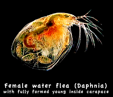
See also Daphnia, Zebra fish and Tadpoles
![]()

![]()