See also ⇒ HERE
...As can be seen in Table 1, the fluoride exposure
brought about a significant decrease in the
testicular zinc concentration and
an increase in lipid…
..
Some species, including humans, have acquired a
spanner somewhere in the genetic works that
prevents them synthesising vitamin C.
Another oft-quoted exception is the
guinea pig, but similar defects are found
in [some] bats, fish, some birds and
many of our closest primate cousins.
Most animals make ascorbic acid to order.
Zinc Protection From Fluoride-Induced
Testicular Injury In The Bank Vole
(Clethrionomys glareolus)
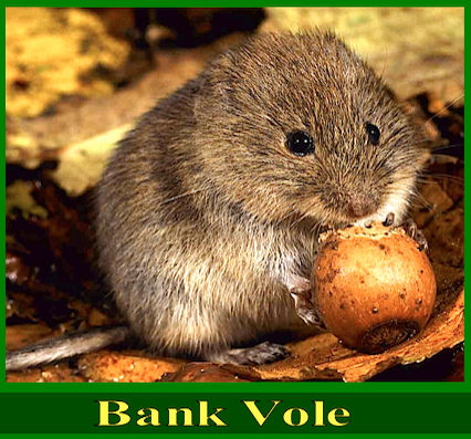
Myodes glareolus
Original ⇒ HERE
Article in Toxicology Letters · April 2004
Impact Factor: 3.26 · DOI: 10.1016/j.toxlet.2003.11.012

Source: PubMed 3 authors, including:
Tadeusz Włostowski University of Bialystok
Poland
32 PUBLICATIONS 372 CITATIONS
SEE PROFILE
Elżbieta Bonda-Ostaszewska
University of Bialystok
15 PUBLICATIONS 230 CITATIONS
.
SEE PROFILE
All in-text references underlined in blue are linked to publications on Research Gate, letting you access and read them immediately.
Available from: Elżbieta Bonda-Ostaszewska
Retrieved on: 24 June 2016
Toxicology Letters 147 (2004) 229–235
Zinc protection from fluoride-induced testicular
injury in The Bank Vole (Clethrionomys glareolus)
Alicja Krasowska∗, Tadeusz Włostowski, El˙zbieta Bonda Institute of Biology, University of Białystok, ´ Swierkowa 20B, 15-950 Białystok, Poland
Received 6 October 2003; received in revised form 30 October 2003; accepted 6 November 2003
.
Abstract:
Previous work has shown that a high fluoride intake in rodents leads to histopathological changes in the germinal epithelium of testes that is associated with zinc deficiency.
The purpose of this study was to determine whether supplemental dietary Zn would protect against testicular toxicity induced by fluoride in a small rodent, the bank vole. The 4-month exposure period to fluoride (200_g/ml of drinking water) induced histopathological changes (hemorrhage in interstitium, necrosis and apoptosis in seminiferous tubule epithelium) which were accompanied by decreased testicular zinc concentration and increased lipid peroxidation.
Supplemental dietary zinc (110–120_g/g) together with fluoride treatment resulted in complete reversal of the fluoride-mediated effects. However, supplemented dietary Zn did not affect the accumulation of fluoride in the testes and bone.
These data suggest that a zinc-enriched diet protects seminiferous tubules against fluoride toxicity by preventing the fluoride-induced testicular zinc deprivation.
© 2003 Elsevier Ireland Ltd. All rights reserved.
Keywords: Fluoride; Zinc; Testes; Histopathology; Apoptosis
.
Introduction
Fluoride (F) is an essential [? * ] trace element which has
a very high affinity for bone mineral where it is incorporated
into the apatite crystal structure by substitution
for hydroxyl ion (Zipkin, 1970). This substitution
confers protective effects against mineral dissolution,
with important implications for animals and human
demineralizing diseases (Guo et al., 1988; Machoy,
1991). However, at higher doses fluoride becomes
toxic and adversely affects a number of physiological
∗ Corresponding author. Fax: +48-857457302.
E-mail address: alak@uwb.edu.pl (A. Krasowska).
processes including reproduction (Weber and Reid,
1969; Messer et al., 1973; Zeiger et al., 1993; Freni,
1994).
It has been suggested that impaired reproduction
may be directly related to an injury of the germinal
epithelium of testicular tubules induced by fluoride
exposure (Kour and Singh, 1980a; Krasowska, 1989).
Previous studies from our laboratory revealed that
testicular necrosis caused by fluoride is accompanied
primarily by a reduction of zinc concentration
in the testes of rats and bank voles (Krasowska and
Włostowski, 1992, 1996). Because a zinc-deficient
diet produces similar histopathological changes in the
germinal epithelium as those brought about by fluoride
exposure (Millar et al., 1958; Mason et al., 1982;
0378-4274/$ – see front matter © 2003 Elsevier Ireland Ltd. All rights reserved.
doi:10.1016/j.toxlet.2003.11.012
230 A. Krasowska et al. / Toxicology Letters 147 (2004) 229–235
Merker and Günther, 1997), it is conceivable that fluoride
affects spermatogenesis through changes in zinc
metabolism. If this is the case, then protection against
fluoride-induced testicular Zn deprivation should prevent
injury to the organ. Therefore, this study was
designed to determine whether supplemental dietary
Zn would protect against histopathological changes
in the testes of bank voles exposed chronically to fluoride.
Since Zn deficiency has been shown to induce
apoptosis and lipid peroxidation in the testes (Oteiza
et al., 1995; Nodera et al., 2001), in the present study
the two processes were also examined.
- .
-
Materials and methods
2.1. Animals and experimental design
Forty male bank voles (1 month old, weighing
11–13 g) from our own laboratory stock were used
throughout the study. The animals were housed in
stainless steel cages (two per cage) under controlled
environmental conditions (18–20 ◦C temperature,
50%–70% relative humidity, 12-h light/dark cycle).
The bank voles were divided into four groups
according to drinking water and dietary zinc: (1)
control—receiving distilled water and food containing
20–25_g Zn/g; (2) fluoride—receiving distilled
water containing 200 _g F/ml as NaF (Krasowska
and Włostowski, 1996) and food containing 20–25 _g
Zn/g; (3) fluoride + zinc—receiving distilled water
containing 200 _g F/ml as NaF and food containing
110–120_g Zn/g, and (4) zinc—receiving distilled
water and food containing 110−120_g Zn/g. For 4
months, bank voles received ad libitum fluoride in
their drinking water and control or zinc-supplemented
wheat grains which are considered to be an adequate
quality food for the bank vole (Włostowski et al.,
1996). The grains supplemented with zinc (soaked in
ZnSO4 solution) and containing 15–20 _g F/g were
prepared prior to the experiment. Atomic absorption
spectrophotometry (AAS) analysis of the grain
revealed that actual levels of Zn were between 20
and 25 _g Zn/g dry weight (control) and 110–120 _g
Zn/g dry weight (Zn-enriched diet). In addition, an
identical amount of apple was offered to all animals
(3 g per vole per week) which ate it completely. The
water intake was measured weekly. The experimental
protocols were approved by the local ethical committee
for performing and experimental study on
laboratory animals (Medical Academy in Białystok).
.
2.2. Assays
At the end of the 4-month exposure period, the bank
voles were weighed, euthanized by cervical dislocation
and both testes and femur were removed. The
left testis was transferred to 2.0 ml chilled 0.25M sucrose
and homogenized with a Teflon pestle in a glass
homogenizer. Aliquots (0.2 and 1.0 ml) of the homogenate
were taken for determination of lipid peroxidation
and zinc concentration, respectively. The
right testis was fixed in Bouin’s fluid. One half of the
testis was processed for histological examination and
immunohistochemistry. The other half was dried and
used for fluoride measurement.
Lipid peroxidation was assessed by measuring
malondialdehyde (MDA) formation, using the thiobarbituric
acid (TBA) assay (Ohkawa et al., 1979).
To 0.2 ml of the tissue homogenate, 0.2 ml of 8.1%
sodium dodecyl sulfate, 1.5 ml of 20% acetic acid,
1.5 ml of 0.8% thiobarbituric acid, and 0.6 ml of distilled
water were added and vortexed. The reaction
mixture was placed in a water bath at 95 ◦C for 1 h.
After cooling, 1.0 ml of distilled water and 5.0 ml
of butanol/pyridine mixture (15:1, v/v) were added
and vortexed. After centrifugation, absorbance
of the organic phase was determined at 532 nm.
Tetraethoxypropane was used to prepare a calibration
curve. The results were expressed as TBARS (nmol/g
wet weight).
Zinc determination was performed as described previously
(Włostowski et al., 1996). Briefly, the homogenate
(1.0 ml) was placed in a glass tube with
2.0 ml of concentrated nitric acid. After 20 h of sample
digestion at room temperature, 72% perchloric
acid (0.5 ml) was added and the mixture was heated
at 100 ◦C for 3 h. Finally, the temperature was raised
to 150 ◦C and digestion continued for another 4 h.
Deionized water was added to the residue after digestion
to a volume of 3.0 ml. Zinc in these solutions
was measured on a flame absorption spectrophotometer
(AAS 3, Zeiss Jena). Samples of bovine liver 1577b
(NIST) were also analyzed in an identical manner to
check accuracy of the method. The recovery of Zn was
90%–95%.
-
Krasowska et al. / Toxicology Letters 147 (2004) 229–235 231
Fluoride determination was performed spectrophotometrically
at 622 nm by using modified lanthanum/
alizarin complexone reagent (Culik, 1986).
The separation of fluoride from dry portion of the
testis (30–35 mg) and bone (6–7 mg) was achieved by
microdiffusion from perchloric acid and absorption
by sodium hydroxide on filter paper in disposable
polypropylene vials.
2.3. Histopathology
One half of each fixed testis was processed by
standard histological methods, stained with hematoxylin
and eosin, and analyzed by light microscopy
for histopathological changes.
2.4. Immunohistochemistry
Apoptosis in the testes was demonstrated in situ by
the TUNEL (TdT-mediated dUTP-fluorescein Nick
End Labeling) assay, using a kit from Roche Diagnostics
(Mannheim, Germany) according to their
instructions. Briefly, sections were dewaxed in xylene,
hydrated in graded alcohol series and permeabilized
in 0.1% Triton X-100/0.1% sodium citrate for
8 min. Terminal deoxynucleotidyl transferase (TdT)
enzyme and fluorescein-labeled nucleotides were applied
to the sections for 60 min at 37 ◦C. Sections
were washed with PBS and treated with alkaline
phosphatase-conjugated anti-fluorescein antiboby for
30 min at 37 ◦C. They were next treated with substrate
solution (NBT/BCIP) for 10 min in dark. Apoptotic
activity was quantified by counting the number of cells
.
Table 1
The effect of fluoride exposure on body and testis weights, testicular zinc and lipid peroxidation (TBARS), testicular and bone fluorideVand testicular injury in the bank vole fed diets supplemented and not supplemented with zinc Control Fluoride Fluoride + Zinc Zinc
Body weight (g) 21.5 °æ 2.8a 22.1 °æ 2.5a 21.7 °æ 2.4a 23.2 °æ 3.0a
Left testis wet weight (mg) 330 °æ 79a 300 °æ 60a 291 °æ 80a 348 °æ 60a
Testicular zinc (_g/g wet weight) 32.5 °æ 3.8a 18.0 °æ 2.0b 31.5 °æ 2.5a 33.0 °æ 2.0a
TBARS (nmol/g wet weight) 52.6 °æ 9.1a 75.0 °æ 11.1b 56.5 °æ 10.6a 51.0 °æ 9.7a
Testicular fluoride (_g/g dry weight) 4.00 °æ 1.50a 11.90 °æ 2.96b 9.00 °æ 1.54b 3.90 °æ 1.35a
Bone fluoride (_g/g dry weight) 247 °æ 38a 4872 °æ 687b 4790 °æ 488b 210 °æ 75a
Apoptosis 0.23 °æ 0.12a 1.16 °æ 0.49b 0.23 °æ 0.09a 0.15 °æ 0.03a
Histopathology − + − −
Values represent the mean °æ S.D. for n = 10. Apoptosis is expressed as TUNEL-positive cells per seminiferous tubule (see Fig. 2).
Histopathology: normal morphology (−), histopathological changes (+) (see Fig. 1). Means in the same row marked with different superscript letters (a and b) are significantly different (P < 0.05). positive for TUNEL staining within entire testis cross section, as proposed by Young et al. (1999). Apoptosis was expressed as number of TUNEL-positive cells per total number of seminiferous tubules within each testis cross.
.
2.5. Statistical analysis
Data were expressed as means °æ S.D. The values
were analyzed by two-way analysis of variance (ANOVA) followed by the Duncan’s multiple-range test (MS Statistica). Differences at P < 0.05 were considered statistically significant.
- .
-
Results
During the 4-month period of observation, fluoride
loading (200_g F/ml) did not affect the consumption
of water, which amounted to 3–4 ml per animal per
day. There were also no significant differences in the
final body and testes weights among the four groups
of bank voles (Table 1).
Histopathological changes (hemorrhage in interstitium,
vacuolization and necrosis of seminiferous epithelium)
(Fig. 1) and increased incidence of apoptosis
(Fig. 2) occurred in the testes of all bank voles exposed
to fluoride alone (Table 1); notably, no lesions
were produced by fluoride in the presence of the extra
dietary Zn.
As can be seen in Table 1, the fluoride exposure
brought about a significant decrease in the
testicular zinc concentration and an increase in lipid
232 A. Krasowska et al. / Toxicology Letters 147 (2004) 229–235
Fig. 1. Representative photomicrographs of testis section from (A) control bank voles and (B, C) bank voles that received water containing 200 _g F/ml (fluoride group) for 4 months. Hemorrhage in interstitium (B) (arrows), and vacuolization and necrosis of the seminiferous epithelium (C) in the fluoride-treated voles were observed. No histopathological changes were seen in bank voles from the control, fluoride + zinc and zinc groups (not shown). H & E staining, 200°ø.
-
Krasowska et al. / Toxicology Letters 147 (2004) 229–235 233
Fig. 2. Immunohistochemical demonstration of apoptotic cells in testes by the TUNEL technique.
(A) Control bank voles showing the normal level of apoptosis.
(B) Bank voles received drinking water containing 200 _g F/ml, for 4 months showing the increased number of apoptotic cells (arrows); 200°ø. peroxidation; supplemental dietary Zn resulted in complete reversal of the fluoride mediated effects.
The two-way analysis of variance revealed that the accumulation of fluoride in the testes and bone of bank voles was significantly affected by the fluoride exposure
(P < 0.0001), but supplemental dietary Zn had no influence on the fluoride concentrations (Table 1).
- .
-
Discussion
Zinc has been shown so far to protect against toxicity of various chemicals (Cagen and Klaassen, 1979;
Miranda et al., 1982; Szyma´nska et al., 1991; Kaji et al., 1993; Zhou et al., 2002). The mechanism of zinc protection has been attributed to the alteration 234 A. Krasowska et al. / Toxicology Letters 147 (2004) 229–235 in pharmacokinetics of xenobiotics, stabilization of membranes, inhibition of P-450-dependent monooxygenase activity, stabilization of cellular thiols or the activation of gluthatione-associated enzymes, inhibition of lipid peroxidation and sequestration of reactive moieties, free radicals and metal ions, by the Zn-induced metallothionein (Chvapil et al., 1972; Cagen and Klaassen, 1979; Goering and Klaassen,
1984; McMillan and Schnell, 1984; Cho and Fong,1990).
The results of the present study indicate that concurrent administration of elevated but physiologic level of dietary Zn can protect seminiferous tubules from the toxic effects of fluoride. This protection was probably not due to an alteration in pharmacokinetics of fluoride, as supplemental dietary Zn did not change the tissue distribution of this element (Table 1). It is thus unlikely that spermatogenesis in the bank vole can be directly affected by fluoride. The assumption in confirmed by results of Sprando et al. (1996) who have demonstrated that spermatogenesis in the adult rat is not adversely affected by direct exposure to fluoride even at levels 200 times greater than those under normal conditions.
The present work showed, however, that supplemental dietary Zn prevents a reduction in the testicular zinc concentration induced by fluoride exposure
(Table 1). The exact mechanism by which fluoride decreases the testicular zinc is unknown at present.
It cannot be ruled out that the decrease may have been due to an inhibition of Zn absorption and/or an increase of its excretion in the urine under the influence of fluoride.
The latter possibility may be involved because kidneys are also adversely affected by prolonged use of NaF (Kour and Singh, 1980b). Moreover, the sequestration of zinc from the plasma by fluorotic bone may also account, at least to some degree, for the deprivation of Zn in the testes as well as in the liver and kidneys (Krasowska and Włostowski, 1992).
Several lines of evidence indicate that reactive oxygen species (ROS) are involved in fluoride-induced impairment of soft-tissue function (Zhi-Zhong et al.,
1989; Rzeuski et al., 1998; Shivarajashankara et al.,
2001). Lipid peroxidation is considered as an indirect
measure of ROS generation (Suzuki et al., 2000).
In the present study, lipid peroxidation increased in the testes of bank voles exhibiting at the same time the tissue injury and apoptosis (Table 1).
Thus, it is possible that fluoride induced oxidative stress could be responsible for these processes. Notably, the fluoride-induced testicular lipid peroxidation, histopathological changes and increased incidence of apoptosis were associated with testicular Zn depletion, and supplemental dietary Zn resulted in complete reversal of the fluoride-mediated effects
(Table 1).
These data suggest that the Zn depletion in testes caused by fluoride exposure may be a causal factor in inducing testicular injury in the bank vole.
The assumption is supported by the fact that both lipid peroxidation and apoptosis in testes have been demonstrated to increase in animals fed Zn-deficient diets (Oteiza et al., 1995; Nodera et al., 2001) and dietary Zn deficiency produces similar histopathological changes in testes as those observed in this study (Millar et al., 1958; Mason et al., 1982; Merker and Günther, 1997). It is also worth noting that both a Zn-deficient diet (Gilabert et al., 1996) and fluoride exposure (Tokar and Savchenko, 1977) induce a reduction in serum testosterone, which in turn can lead to an inhibition of spermatogenesis, further supporting the notion that zinc plays an important role in the pathogenesis of fluoride-induced testicular injury.
.
In conclusion, the results from the present study demonstrate that a zinc-enriched diet protects seminiferous tubules against fluoride toxicity.
This protection appears to be due to the prevention of fluoride-induced testicular zinc deprivation.
.
References:
Cagen, S.Z., Klaassen, C.D., 1979. Protection of carbon tetrachloride- induced hepatotoxicity by zinc: role of metallothionein.
Toxicol. Appl. Pharmacol. 51, 107–116.
Cho, C.H., Fong, L.Y., 1990. The interaction of ethanol and zinc on hepatic glutathione and glutathione transferase activity in mice. Agents Actions 29, 328–385.
Chvapil, M., Ryan, J.N., Zukowski, C.F., 1972. Effect of zinc on lipid peroxidation in liver microsome and mitochondria. Proc.
Soc. Exp. Biol. Med. 141, 150–153.
Culik, B., 1986. Microdiffusion and spectrophotometric determination of fluoride in biological samples. Anal. Chim. Acta 189, 329–337.
Freni, S.C., 1994. Exposure to high fluoride concentrations in drinking water is associated with decreased birth rates. J. Toxicol. Environ. Health 42, 109–121.
Krasowska et al. / Toxicology Letters 147 (2004) 229–235 235
Gilabert, E.R., Ruiz, E., Osorio, C., Ortega, E., 1996. Effect of dietary zinc deficiency on reproductive function in male rats: biochemical and morphometric parameters. J. Nutr. Biochem.
7, 403–407.
Goering, P.L., Klaassen, C.D., 1984. Zinc-induced tolerance to
cadmium hepatotoxicity. Toxicol. Appl. Pharmacol. 74, 299–307.
Guo, M.K., Nopakun, J., Messer, H.H., Ophang, R., Singer, L.,
-
Retention of skeletal fluoride during bone turnover in rats. J. Nutr. 118, 362–366.
Kaji, T., Mishima, A., Yamamoto, C., Sakamoto, M., Kozuka, H., Zinc protection against cadmium-induced destruction of the monolayer of cultured vascular endothelial cells. Toxicol.
Lett. 66, 247–255.
Kour, K., Singh, J., 1980a. Histological finding of mice testes following fluoride ingestion. Fluoride 13, 160–162.
Kour, K., Singh, J., 1980b. Histological findings in kidneys of mice following fluoride administration. Fluoride 13, 163–167. Krasowska, A., 1989. Influence of low-chitin krill meal on reproduction of Clethrionomys glareolus (Schreber, 1780).
Comp. Biochem. Physiol. C 94, 313–320. Krasowska, A., Włostowski, T., 1992. The effect of high fluoride intake on tissue trace elements and histology of testicular tubules in the rat. Comp. Biochem. Physiol. C 103, 31–34.
Krasowska, A., Włostowski, T., 1996. Photoperiodic elevation of testicular zinc protects seminiferous tubules against fluoride toxicity in the bank vole (Clethrionomys glareolus).
Comp. Biochem. Physiol. C 113, 81–84.
Machoy, Z., 1991. Are the fluoride content and the physical load of bones in man related. Fluoride 24, 100–102.
Mason, K.E., Burns, W.A., Smith, J.C., 1982. Testicular damage associated with zinc deficiency in pre- and post-pubertal rats: response to zinc repletion. J. Nutr. 112, 1019–1028.
McMillan, D.A., Schnell, R.C., 1984. Zinc protection against
bromobenzene induced hepatotoxicity in the rat. Toxicologist
4, 451.
Merker, H.J., Günther, T., 1997. Testis damage by zinc deficiency
in rats. J. Trace Elem. Med. Biol. 11, 19–22.
Messer, H.H., Armstrong, W.D., Singer, L., 1973. Influence of
fluoride intake on reproduction in mice. J. Nutr. 103, 1319–1326.
Millar, M.J., Fischer, M.J., Elcoate, P.V., Mawson, C.A., 1958. The
effect of dietary zinc deficiency on the reproductive system of
male rats. Can. J. Biochem. Physiol. 36, 557–569.
Miranda, C.L., Henderson, M.C., Reed, R.L., Schmitze, J.A.,
Buhler, D.R., 1982. Protective action of zinc against
pyrrolizidine alkaloid-induced hepatotoxicity in rats. J. Toxicol.
Environ. Health 9, 359–366.
Nodera, M., Yanagisawa, H., Wada, O., 2001. Increased apoptosis
in a variety of tissues of zinc-defficient rats. Life Sci. 69,
1639–1649.
Ohkawa, H., Ohishi, N., Yagi, K., 1979. Assay for lipid
peroxides in animal tissues by thiobarbituric acid reaction.
Anal. Biochem. 95, 351–358.
Oteiza, P.I., Olin, K.L., Fraga, C.G., Keen, C.L., 1995. Zinc
deficiency causes oxidative damage to proteins, lipids and DNA
in rat testes. J. Nutr. 125, 823–829.
Rzeuski, R., Chlubek, D., Machoy, Z., 1998. Interactions between
fluoride and biological free radical reactions. Fluoride 31, 43–
45.
Shivarajashankara, Y.M., Shivashankara, A.R., Gopalakrishna-
Bhat, P., Hanumanth-Rao, S., 2001. Effect of fluoride
intoxication on lipid peroxidation and antioxidant systems in
rats. Fluoride 34, 108–113.
Sprando, R.L., Black, T.N., Ames, M.J., Rorie, J.I., Collins, T.F.X.,
-
Effect of intratesticular injection of sodium fluoride on
spermatogenesis. Food Chem. Toxicol. 34, 377–384.
Suzuki, Y., Apostolova, M.D., Cherian, M.G., 2000. Astrocyte
cultures from transgenic mice to study the role of
metallothionein in cytotoxicity of test-butyl hydroperoxide.
Toxicology 145, 51–62.
Szyma´nska, J.A., ´ Swietlicka, E.A., Piotrowski, J.K., 1991.
Protective effect of zinc in the hepatotoxicity of bromobenzene
and acetaminophen. Toxicology 66, 81–91.
Tokar, V.I., Savchenko, O.N., 1977. The influence of inorganic
fluorine compounds on functional condition of the hypophysistestis
system. Probl. Endokrinol. 23, 104–107.
Weber, C.D., Reid, B.L., 1969. Fluoride toxicity in the mouse. J.
Nutr. 97, 90–94.
Włostowski, T., Krasowska, A., Dworakowski, W., 1996. Low
ambient temperature decreases cadmium accumulation in the
liver and kidneys of the bank voles (Clethrionomys glareolus).
Biol. Metals 9, 363–369.
Young, K.A., Zirkin, B.R., Nelson, R.J., 1999. Short photoperiod
induces testicular apoptosis in the white-footed mouse
(Peromyscus leucopus). Endocrinology 140, 1331–1339.
Zeiger, E., Shelby, H.D., Witt, K.L., 1993. Genetic toxicity of
fluoride. Environ. Mol. Mutagen. 21, 309–318.
Zhi-Zhong, G., Pei-Si, Y., Nai-den, Y., Zong-Jie, Z., 1989. An
experimental study of blood biochemical diagnostic indices for
chronic fluorosis. Fluoride 22, 112–118.
Zhou, Z., Sun, X., Lambert, J.C., Saori, J.T., Kang, Y.J., 2002.
Metallothionein-independent zinc protection from alcoholic
liver injury. Am. J. Pathol. 160, 2267–2274.
Zipkin, I., 1970. Physiological effects of small doses of fluoride:
effects on the skeleton of man. WHO Monograph Ser. 59, 185–201.
[? * ] F. is a serious environmental pollutant – we need to reduce our intake – and stop contaminating our water supply with it.
This research is therefore significant.
We thank you for this paper.
Note:
Since then a few species,
including humans, have acquired
a spanner somewhere in the genetic
works that prevents them synthesising
vitamin C. Another oft-quoted exception is the
guinea pig, but similar defects are found in bats, fish,
birds and many of our closest primate cousins.
Most animals make ascorbic acid to order.
Birds are known to synthesize vitamin C
in sufficient amounts, many feel it
is not necessary in their diets.
We have noticed that in times of stress,
and that includes at breeding times,
our birds consume larger amounts of
foods containing this vitamin;
thus, we feel it to be especially
useful at these times.

![]()